
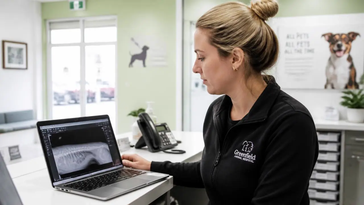
Every practice sends its first study to an outside radiologist a little differently: a frantic Friday-night GDV that cannot wait for the local specialist, a slow-growing thoracic mass that the referring vet wants a second opinion on, an ultrasound the associate is not confident reading alone. What almost nobody does is sit down beforehand and learn the small set of things that determine whether that first experience goes well or badly. This is that sitting-down.
Key takeaways
- A teleradiology report is a specialist opinion built from the data you send. It cannot exceed the quality of that data.
- Full DICOM, not a JPEG export and never a phone photo of a monitor, is the difference between a confident report and a hedged one.
- Clinical history is the single cheapest, highest-leverage input a practice controls. A blank field costs you accuracy for free.
- Pricing models are not interchangeable. Match per-study, per-modality or subscription pricing to your actual monthly caseload.
- Board certification is a floor, not a differentiator. Coverage, communication and QA process separate providers who clear that floor.
- Treat the first 90 days as a rollout with owners, not a switch you flip. Most failures are workflow failures, not radiology failures.
What a teleradiology report is, and is not
A teleradiology report is a written interpretation of imaging data by a radiologist who is not physically present in your building, produced from the same DICOM images your local specialist would look at if one were available. It is not a diagnosis handed down in isolation, and it is not a substitute for the physical exam, bloodwork or clinical judgment that only the veterinarian in the room can supply. The report is one input into a case, and it is only as good as the study, the history, and the question it is asked to answer.
It also is not instantaneous magic. A radiologist reading remotely goes through the same systematic search a radiologist reading in-house would: survey the whole study, work through each organ system or region methodically, then form an impression tied to the findings. What teleradiology changes is who does that work and when, not the nature of the work itself.
What you should expect to receive
- A findings section describing what was actually seen, with measurements and laterality where relevant.
- An impression that ranks differentials against the findings rather than restating them.
- Explicit limitations: what the study cannot rule out, and why.
- A specific recommendation, not a generic instruction to correlate clinically.
DICOM versus JPEG, and why phone photos fail
DICOM is not a file format in the way JPEG is. It is a full data structure that carries the pixel data at its original resolution and bit depth, along with acquisition parameters, patient and study metadata, and, critically, the ability to be windowed and leveled after the fact. A JPEG export flattens all of that into a single fixed-contrast image. Once it is flattened, no radiologist on earth can recover the soft tissue detail or the subtle bone lesion that a different window level would have revealed.
A phone photo of a monitor compounds the problem: screen glare, moire patterns from the monitor's pixel grid, ambient lighting, and the monitor's own display calibration are now baked permanently into the image, on top of the JPEG compression it was already going to suffer if exported directly. This is not a minor quality complaint. It is the difference between a radiologist who can confidently characterize a lesion margin and one who has to write around a fundamental unknown.
| Format | Windowing possible | Typical failure mode in the report |
|---|---|---|
| Native DICOM | Yes, full range | None specific to format |
| Exported JPEG or PNG | No, fixed at export settings | Subtle findings at the extremes of the gray scale are missed or hedged |
| Screenshot of a workstation | No | Compression artifact mistaken for or masking real pathology |
| Phone photo of a monitor | No | Glare, moire and calibration loss; report frequently recommends a repeat study |
What each modality needs to be read well
Every modality has its own version of the DICOM problem: a specific way the data needs to arrive for the radiologist to do the job you are paying for.
Digital and computed radiography (DR/CR)
Send the complete series for the region and clinical question, including orthogonal views. A single oblique view sent because it "looked interesting" is a request for a hedge, not a diagnosis. Label laterality clearly and consistently; mislabeled left and right is a small error that produces a materially wrong report.
Ultrasound: clips versus stills
A still image shows a radiologist one frame of a moving, three-dimensional structure. A cine clip shows motion, which is often how a mass is distinguished from a normal loop of bowel, how a mobile foreign body is identified, or how cardiac wall motion is assessed. Send clips whenever your machine can generate them, not just the frozen frames you happened to save. If your protocol only ever saves stills, that is worth revisiting with whoever trained your sonographer.
CT series and reconstructions
Send the full raw series, not just the reconstructed images your console displayed after the scan. Radiologists frequently need to reconstruct in a different plane, slice thickness or algorithm than the one your console defaulted to, and that is only possible if the raw data made the trip. Contrast timing and phase should be noted in the history, since a non-contrast, arterial and portal-venous phase image can look similar at a glance but mean very different things.
MRI sequences
Send every sequence acquired, not a curated subset. T1, T2, STIR or fat-saturated sequences, and post-contrast series each answer a different question, and a lesion that is inconspicuous on one sequence can be obvious on another. Omitting sequences to save transfer time is a false economy that shows up as a less specific differential list.
The clinical history field is your biggest lever
If there is one habit that separates practices who get consistently strong reports from practices who get consistently hedged ones, it is what gets typed into the history field. "Vomiting" tells a radiologist almost nothing. "Six-year-old intact male Labrador, acute vomiting for 18 hours, painful cranial abdomen on palpation, known fabric ingestion two days ago" tells the radiologist exactly what to hunt for and how urgently to hunt for it.
- Signalment. Species, breed, age and sex change the differential list before a single image is opened.
- Duration and progression. Acute versus chronic changes which differentials are even plausible.
- Relevant exam findings. Pain location, palpable masses, auscultation findings.
- Medications and recent procedures. Steroids, chemotherapy, recent surgery all alter expected imaging appearance.
- Prior imaging. Attach it or name it. A radiologist comparing to a study they cannot see cannot comment on progression.
- A specific question. "Assess for mechanical obstruction" produces a sharper report than "please evaluate."
The turnaround versus accuracy piece covers this from the speed side: a complete history is also one of the few genuine ways to make a report faster without cutting corners, because the radiologist is not spending time guessing at context you already had in the exam room.
Free for veterinary teams
The first-study submission checklist
A one-page checklist your front desk and technicians can keep by the imaging workstation: what to export, what to type into history, and how to flag STAT correctly. Ask and we will send it.
- Modality-by-modality export checklist
- A fill-in-the-blank clinical history template
- The STAT criteria we recommend posting by the console
No contracts. No minimums. Radiologists on shift every day of the year.
STAT versus routine, and how to choose
STAT should mean a decision in your building is actively waiting on the read. A one-hour target for STAT and a 24-hour target for routine, available every day of the year, is a realistic working standard that a provider offering AVMA-aligned emergency and specialty care should be able to meet. Flagging everything as STAT does not make your practice faster; it makes your STAT queue behave like a routine queue, which defeats the purpose for the case that actually needs it.
- A patient in visible distress where surgery timing depends on the read.
- Suspected GDV, obstruction, or acute abdomen where the next step is decided by the finding.
- A trauma case where the extent of injury changes whether the patient is stabilized or referred immediately.
Everything else, including most staging studies, wellness screening and pre-anesthetic imaging, belongs in routine. Routine within 24 hours is fast enough medicine for the vast majority of cases, and it keeps STAT meaningful for the cases where an hour actually changes the outcome.
Pricing models and the hidden costs
Teleradiology pricing is not standardized across the industry, and the model that looks cheapest on a rate sheet is not always the cheapest for your actual caseload.
| Model | How it works | Fits best when | Watch for |
|---|---|---|---|
| Per study | Flat rate regardless of modality | Low, unpredictable monthly volume | May undervalue complex CT/MRI reads, pushing providers to rush them |
| Per modality | Separate rate for radiographs, ultrasound, CT, MRI | Mixed caseload with a few high-complexity studies | Rate card complexity; ask for the full list up front |
| Subscription | Fixed monthly fee for a volume or unlimited reads | Consistent, higher monthly volume, multi-doctor practices | Overpaying in slow months, or overage fees once you exceed the bundle |
| Minimums or contracts | Guaranteed monthly spend or term commitment | Rarely favors the practice | A provider that needs a minimum is managing its risk with your money |
Hidden costs tend to cluster around three things: STAT surcharges that are not disclosed until the invoice arrives, per-image or per-series charges layered on top of a quoted per-study price, and cancellation or minimum-volume penalties buried in a contract. Ask for a sample invoice before you commit, not just a rate sheet.
Credentialing: what DACVR and ECVDI actually mean
A radiologist certified as a Diplomate of the American College of Veterinary Radiologists (DACVR), or the European equivalent through the European College of Veterinary Diagnostic Imaging (ECVDI), has completed a multi-year residency in veterinary imaging beyond general veterinary training, and passed a rigorous board examination, and maintains ongoing continuing education to keep the credential current. This is what "board-certified" means when a provider uses the phrase, and it is worth confirming rather than assuming, since the term gets used loosely in marketing.
Certification is the floor every serious provider clears, not the feature that separates one provider from another. Ask who specifically reads your studies, whether that person is reachable if you have a question, and how the provider handles peer review and disagreement. The case for a second reader goes deeper into why a documented QA process matters as much as the credential itself.
Legal and record-keeping considerations
A teleradiology report becomes part of the patient's permanent medical record the moment it is issued, and your state veterinary board's record retention rules apply to it exactly as they apply to any other diagnostic finding. Practices sometimes assume the provider's portal will serve as a permanent archive; do not rely on that. Confirm reports and the associated images are pulled into your own PACS or practice management system, so your records survive a provider switch, a service outage, or a portal access change.
- Confirm who holds the legal patient record: your practice, not the provider, in nearly every jurisdiction.
- Verify your AAHA-aligned record-keeping standards are met for imaging reports the same way they are for in-house diagnostics.
- Ask what happens to your studies if you stop using a provider: export access, deletion timelines, and format.
- Keep a written note of the specific radiologist and date for every report, in case a second opinion or a legal question arises later.
A first-90-days rollout plan
Most teleradiology disappointments in the first few months trace back to workflow, not radiology. A short, deliberate rollout avoids nearly all of them.
Days 1 to 14: setup and one test case
- Confirm DICOM export or send is configured correctly on every modality you plan to use.
- Post the STAT criteria by every imaging workstation, in plain language.
- Build a clinical history template your team can fill in under 60 seconds.
- Send one low-stakes case as a test before your first true emergency depends on the workflow working.
Days 15 to 45: the whole team, and the exceptions
- Train every shift, not just the shift that happened to be present at rollout, including relief staff.
- Run one drill for the DICOM transfer failing, so someone knows the fallback before it happens for real at 2am.
- Review the first ten reports as a team against the quality markers above.
Days 46 to 90: measure and adjust
- Pull actual turnaround data for your studies and compare it to what was promised.
- Ask your provider for their peer review and escalation process in writing if you have not already.
- Decide, with real data, whether your pricing model still matches your monthly volume.
None of this needs to be complicated, and it does not need a contract to start. If you want to talk through what a rollout would look like for your specific caseload and modalities, reach out or read more about how our radiologists work before you send the first study.
Frequently asked questions
Work with us
Board-certified reads, 1 hour STAT, every day of the year
No contracts, no minimums, and direct access to the radiologist reading your case.