
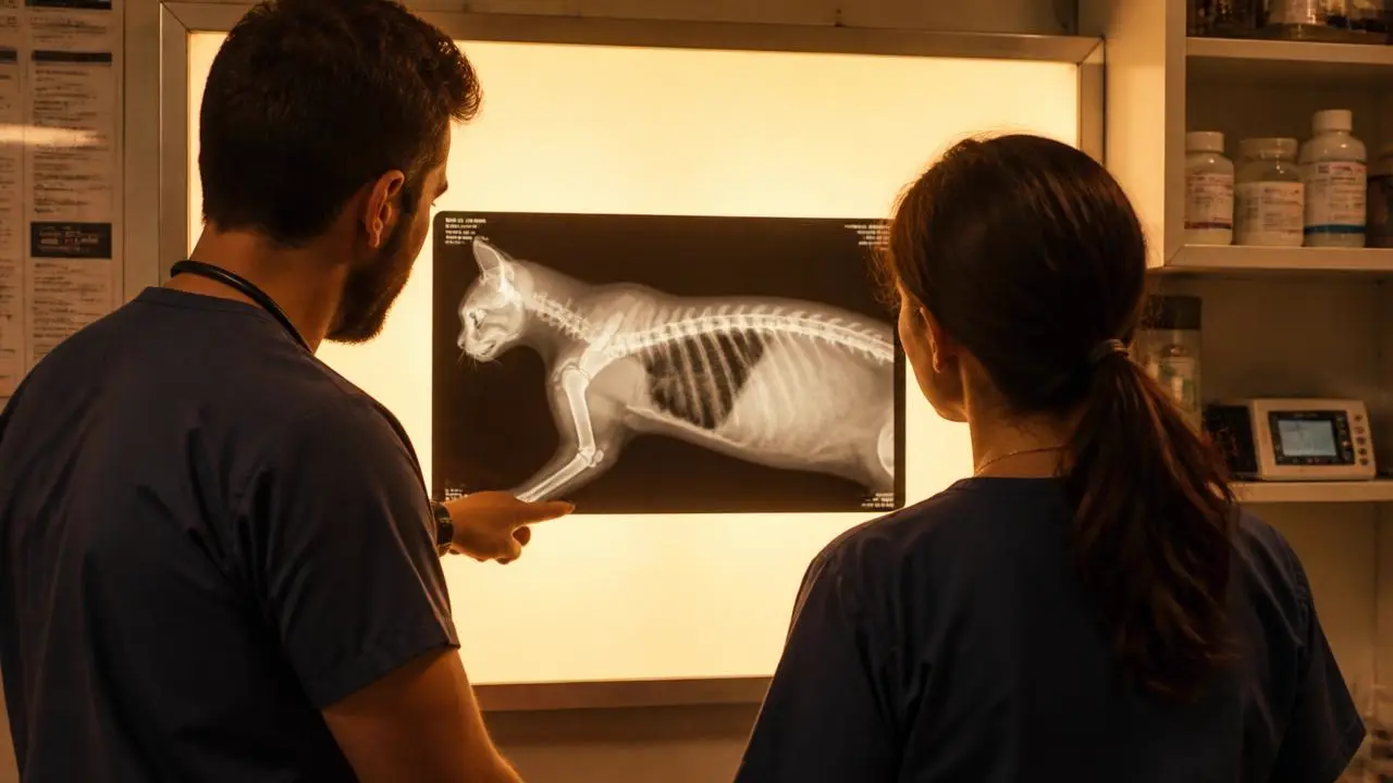
There is a particular kind of resistance to sending studies out, and it is not really about cost. It is the quiet sense that a good clinician ought to be able to read their own films. Most experienced practitioners can read most of their own films. That is not the argument.
The argument is that image interpretation has four specific failure modes that get worse with experience, workload and confidence, not better. Understanding them is the difference between using a second reader as a crutch and using one as an instrument.
Key takeaways
- Satisfaction of search: finding one abnormality measurably reduces the chance of finding a second, in experts as much as novices.
- A clinical question focuses attention, which is useful for the question and dangerous for everything outside it.
- Reading conditions in practice, ambient light, a consult-room monitor, a two-minute window between appointments, degrade detection independent of skill.
- A written, dated specialist report with explicit limitations is a materially stronger record than a clinician one-liner.
- The point of a second read is not correction. It is the second complete search you were never going to perform yourself.
This is not a competence question
Screening programmes in human medicine adopted double reading not because the first radiologist was inadequate, but because two independent complete searches of the same image find more than one search does. The effect is structural. It comes from how visual search works, from how attention narrows around a task, and from the fact that a second reader has not already formed a hypothesis about the case.
In veterinary practice the asymmetry is larger, because the first reader is usually also the clinician who took the history, palpated the abdomen, positioned the patient and is about to quote the owner. All of that context helps. All of it also primes.
1. Satisfaction of search
The most reliably reproduced finding in the radiology perception literature is this: once a reader detects an abnormality, their probability of detecting a second, unrelated abnormality on the same image drops. Not because they stop caring, but because the search task feels resolved.
In small animal practice this looks like:
- The obvious mid-shaft fracture on the pelvic film, and the sacroiliac luxation nobody mentioned.
- The impressive gastric foreign body, and the interstitial nodule at the edge of the caudal lung lobe.
- The confirmed pneumonia, and the mildly enlarged sternal lymph node that would have reframed the whole case.
- The dramatic finding you called correctly at 2am, and the second finding on the same film that was never searched for because the case had already been solved.
2. Anchoring on the clinical question
A specific clinical question sharpens a report. It is also a spotlight, and a spotlight makes everything outside it darker. "Assess for mechanical obstruction" gets you a rigorous assessment of bowel populations and a much weaker assessment of the sublumbar region, the prostate and the caudal lung lobes that crept onto the film.
This is why a good radiology report has two sections: findings and impression. The findings section is an inventory that exists independently of your question. It is where the incidental adrenal nodule, the vertebral endplate lysis, and the healing rib fracture in a cat with a vague history get written down. Those are the findings a second reader is structurally more likely to produce, because they arrived at the study without your hypothesis.
Free for veterinary teams
The second-reader triage card
A one-page decision card for the treatment room: which studies genuinely warrant a specialist read, which do not, and what to include so the report answers the question you actually have. Email us and we will send it over.
- Send / do not send criteria by presentation
- The six history fields that most change a report
- STAT versus routine, so your urgent queue means something
No contracts. No minimums. Radiologists on shift every day of the year.
3. The conditions you read under
Detection of low-contrast lesions is measurably sensitive to display quality, ambient lighting and viewing time. Those are not abstract variables. In practice, the first read of a study frequently happens:
- On a consult-room monitor calibrated for spreadsheets, in a room with the lights on.
- In the ninety seconds between a discharge and the next appointment.
- With the owner standing beside you, watching your face.
- At the end of a twelve-hour shift, on your fourth emergency.
A radiologist reads on calibrated diagnostic displays, in a dark room, with the full DICOM dataset and the ability to window and level, and without an owner in the room. The difference in output is not a difference in ability. It is a difference in conditions, and conditions are much easier to fix by routing the study than by renovating the practice.
| Variable | Clinician first read | Specialist second read |
|---|---|---|
| Display | Office-grade monitor, ambient light | Calibrated diagnostic display, controlled lighting |
| Data | Often a viewer preview or exported image | Full DICOM with window and level control |
| Time on study | Seconds to a couple of minutes | Uninterrupted, proportional to study complexity |
| Prior hypothesis | Strong, formed in the exam room | None, arrives at the images cold |
| Search pattern | Task-directed at the clinical question | Systematic inventory of the whole study |
4. The record, and what it has to survive
Records are read later, by people who were not there: a referral specialist, a covering colleague at 3am, an insurer, occasionally a regulator. "Chest rads NAD" is a note. A dated report that lists what was examined, what was normal, what was abnormal, and explicitly what the study could not exclude is a document.
The clinically useful part is the stated limitation. "No pulmonary nodules identified; radiographic sensitivity for nodules under approximately 5 to 7mm is limited and CT is recommended if exclusion of metastasis is required" tells the next reader precisely how much weight this study can carry. That sentence is worth more to a future colleague than the entire normal findings list. We covered the substance of that particular limitation in the guide to pulmonary nodules and their mimics.
What actually changes in the report
In practice, specialist overreads tend to change cases in four ways, roughly in order of frequency:
- Added incidental findings that alter the anaesthetic plan, the staging discussion, or the recheck interval.
- Downgrades. A great deal of value sits in "this is a pulmonary osteoma, not a metastasis" and "this is an end-on vessel". Preventing an unnecessary oncology referral is as valuable as catching a tumour.
- Reframing the differential rather than adding a finding: the same opacities, a different pattern description, a different disease list.
- Outright missed lesions, which are the rarest and the most talked about.
When to send, and when not to bother
| Situation | Send? | Reason |
|---|---|---|
| The read determines whether you operate tonight | Yes, STAT | The decision is the product, not the image |
| Cancer staging or exclusion | Yes | Stated limitations matter as much as findings |
| Any CT or MRI study | Yes | Cross-sectional interpretation is specialist work by volume alone |
| Images and clinical picture disagree | Yes | The disagreement is usually where the diagnosis is hiding |
| Ultrasound performed by a non-specialist | Usually | Operator-dependent, and clips can be reviewed independently |
| Obvious mid-shaft fracture, clean, single injury | Often not | The whole study should still be reviewed, but a report may add little |
| Recheck confirming expected healing | Often not | Comparison against a prior read is the value, not a new opinion |
Making a second read routine, not exceptional
Practices that get value from a second reader do three things consistently:
- They write a criterion into the protocol, so that sending is a workflow rule rather than a per-case judgement about one's own ability. Nobody has to feel anything about it.
- They submit a real clinical question with real history, because a report is only as good as the context it was written against.
- They call the radiologist on the ambiguous ones. Five minutes on the phone resolves more than three report addenda, and it should not require an escalation process to get through.
The professional literature on diagnostic error, from the AVMA to the human radiology perception research it borrows from, keeps landing in the same place: expertise does not remove these effects, it just makes them less visible to the expert. That is precisely why the second pair of eyes should not be the exception in your workflow.
If you want to see what that looks like on your own caseload, send us one study you have already read and compare the reports. Our modality selection guide is a good companion for deciding what to send in the first place, and you can reach a radiologist directly any hour of any day.
Frequently asked questions
Work with us
Board-certified reads, 1 hour STAT, every day of the year
No contracts, no minimums, and direct access to the radiologist reading your case.