
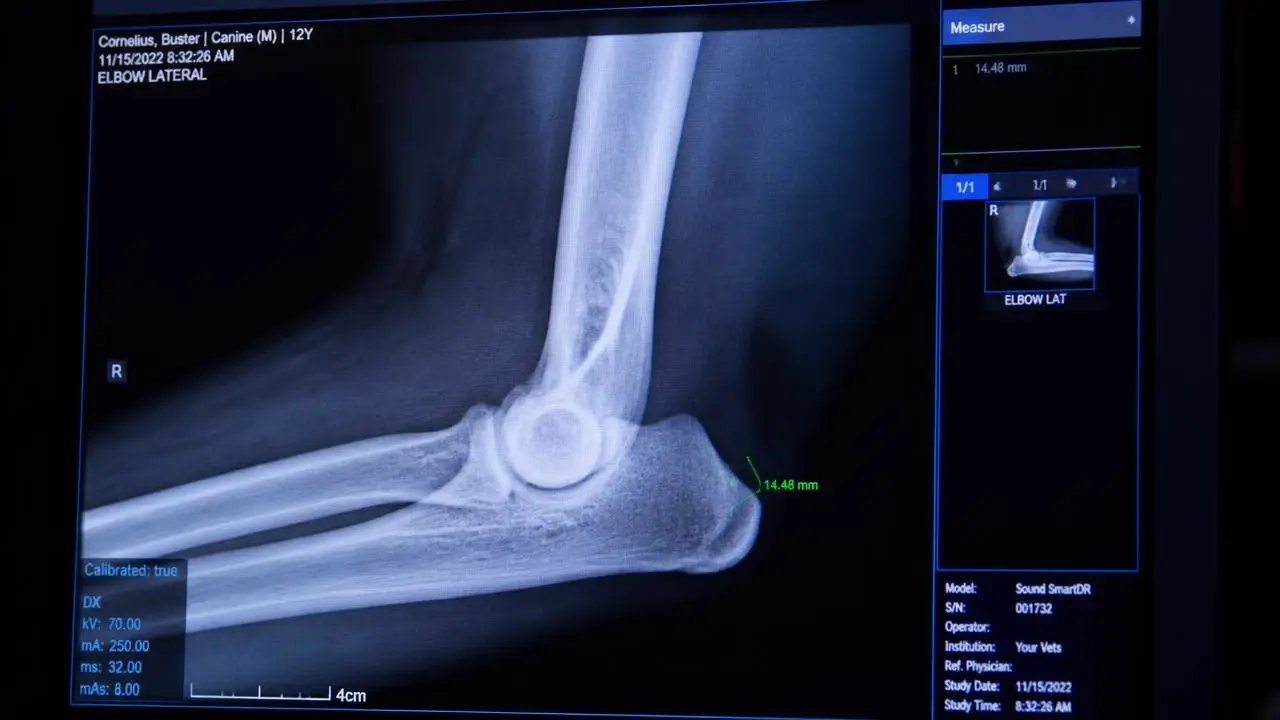
AI has entered veterinary imaging quietly, mostly as a feature inside software you already use rather than a product you decided to buy. A vertebral heart score gets drawn automatically. A study gets flagged as possibly abnormal before anyone opens it. A positioning warning pops up before the plate is even sent. None of that is nothing, and none of it is a radiologist. The gap between those two sentences is where most of the confusion in this space lives.
Key takeaways
- Current veterinary AI tools are narrow: triage flags, automated measurements, positioning checks. None of them read a study the way a radiologist does.
- Training data is weighted toward common breeds and typical conformations, so performance on brachycephalics, chondrodystrophic breeds, cats and exotics is often unverified.
- False positives are not free. Every flag that goes to a client costs a phone call, an anxious owner, and sometimes an unnecessary recheck.
- Automation bias is a real risk: a normal AI screen can make a reader look less carefully, and a positive flag can anchor attention on one finding.
- The strongest evidence favors AI as a radiologist's internal aid, not as a standalone substitute a general practice interprets on its own.
What current veterinary AI tools actually do
Strip away the vendor language and the tools on the market in 2026 fall into a small number of functional categories:
- Triage and worklist flags. A model scores a study as likely normal or possibly abnormal, and the queue reorders so the flagged study gets seen sooner. This is the most defensible use case because the downside of a wrong flag is a delay, not a decision.
- Measurement automation. Vertebral heart score, cardiac silhouette tracing, some joint angle measurements. These replace a ruler and a steady hand, not clinical judgement, and they are only as good as the landmark points the model chooses.
- Positioning and technique checks. Software that flags rotation, under-exposure, or a cut-off field of view before the study is submitted, which is a genuinely useful quality control step that has nothing to do with diagnosis.
- Pattern-specific hit lists. Narrow detectors trained on one finding type, commonly pulmonary nodules, certain fractures, or urinary calculi, that produce a bounding box or a probability score for that one thing and nothing else.
Notice what is absent from that list: nothing on the market synthesizes an entire study, integrates clinical history, or produces a ranked differential diagnosis the way a report does. That is not a temporary gap waiting on the next software version. It reflects how these models are built, one narrow task at a time, trained on labeled examples of that task.
What the marketing claims versus what ships
Vendor materials tend to describe capability in aggregate terms, "detects abnormalities with high accuracy," without specifying which abnormalities, on which body region, in which species, at what threshold. Three gaps recur:
- Scope creep in the pitch, narrow scope in the product. A tool validated on thoracic radiographs for one or two specific findings gets marketed with language that implies general screening capability.
- Performance figures from a curated dataset, not from the messy case mix a general practice actually submits: poor positioning, obese patients, motion artifact, concurrent disease.
- Silence on the denominator. A sensitivity figure without the corresponding false positive rate tells you almost nothing about whether the tool is useful in practice.
The training data problem
Veterinary imaging datasets are smaller and messier than their human medicine equivalents, and they are not evenly distributed across the population a model will eventually be used on.
Species and breed skew
Most publicly available and vendor-assembled training sets are dog-heavy, and within dogs, weighted toward common breeds seen at teaching hospitals and large referral centers. Cats, exotics, and less common breeds are thin in most datasets, and a model's confidence score does not decline to reflect that thinness. It will produce a number either way.
Body shape variation
Chondrodystrophic breeds, deep-chested breeds, brachycephalics and markedly obese or markedly thin patients all present different silhouette geometry on plain films. A cardiac silhouette or VHS tool calibrated on a training set of mixed medium-breed dogs may draw landmark points that do not generalize cleanly to a Dachshund or a Bulldog, and the error will not announce itself.
Label quality
A model is only as good as the ground truth it was trained against. If the labels came from a single reader's impressions rather than a consensus or a confirmed diagnosis, the model has learned that reader's pattern, including their blind spots.
Veterinary schools and colleges publishing imaging research, including groups affiliated with the American College of Veterinary Radiology, have flagged dataset diversity as an open problem rather than a solved one. That caution is worth taking seriously before a tool is trusted on a patient outside its training distribution.
The false positive burden and the client conversation cost
A flag is not free once it leaves the software. Every positive triggers a decision: call the owner, recommend a recheck, order an additional view, or override the flag and document why. None of those options cost nothing.
| Cost type | What it looks like | Who absorbs it |
|---|---|---|
| Clinical time | Reviewing and overriding a flag that a radiologist would have dismissed in seconds | The reading radiologist or the GP interpreting alone |
| Client relationship | An anxious phone call about a finding that resolves to normal on review | Front desk and the attending veterinarian |
| Financial | An unnecessary recheck study or referral triggered by an unconfirmed flag | The client, and eventually the practice's reputation |
| Trust erosion | Repeated false flags train staff to ignore the tool, undermining the genuine catches | The practice's workflow long term |
A tool tuned for high sensitivity to avoid missing anything will, by construction, generate more false positives. That tradeoff is a legitimate design choice, but it is a choice, and a practice adopting the tool should know which way it was tuned and be prepared for the conversation volume that comes with it.
Free for veterinary teams
AI vendor evaluation checklist
A one-page checklist for evaluating any AI feature attached to a radiology or teleradiology product, including the specific questions on training data, false positive rates, and human review that vendors are least eager to answer in a sales call.
- 10 questions to ask before adopting an AI-assisted imaging tool
- What a defensible performance breakdown looks like versus a marketing number
- How to structure a 90-day internal trial before committing
No contracts. No minimums. Radiologists on shift every day of the year.
Automation bias in the reading room
Automation bias is well documented in human radiology and there is no reason to expect veterinary readers are immune to it. It shows up in two directions:
- Under-reading a normal flag. If the software says a study is likely normal, a reader under time pressure may give it a lighter look than they would otherwise, which is exactly backward, since the AI's confidence in "normal" is not equivalent to a radiologist's systematic search.
- Anchoring on a positive flag. A bounding box around one finding can pull attention there and away from a second, unrelated finding elsewhere in the same study, the classic satisfaction-of-search error, now with a visual cue reinforcing it.
The mitigation is procedural, not technological: a reader completes their own systematic search of the full study before consulting the AI output, not after. Reversing that order is a small workflow decision with a real effect on what gets missed. This is one reason the case for a second reader on ambiguous studies does not go away just because software is also looking at the image. A second qualified human reader and an AI flag are not interchangeable safeguards.
Where AI plus a radiologist beats either alone
The strongest evidence for veterinary imaging AI is not AI replacing interpretation, it is AI narrowing a radiologist's attention or automating a measurement so the radiologist's time goes toward judgement rather than mechanics.
- Triage under volume. A radiologist working through an overnight queue benefits from a defensible re-ordering that surfaces a possibly urgent study sooner, provided the STAT process does not depend on the flag being correct.
- Consistent measurement. Automated VHS or similar measurements remove inter-observer variability on the mechanical part of the task, freeing the radiologist to focus on whether the measurement fits the clinical picture.
- A pre-read second look. Used as a checklist prompt after the radiologist's own search is complete, a narrow detector can catch the rare miss the way a checklist catches a rare omission, without displacing the primary read.
Used this way, AI is an instrument in an experienced hand, comparable to how a good queue design and staffing model buys speed without borrowing from accuracy. Used the other way, as a stand-in for the hand, it inherits every one of its narrow-training limitations with no one positioned to catch them.
Regulatory and liability position
Human medicine AI tools intended for diagnostic use generally move through an FDA clearance pathway that requires submitted performance data against a defined use case. Veterinary devices and software largely fall outside that kind of premarket review. A veterinary AI tool can ship and be marketed with far less external validation than its human medicine counterpart, and the burden of proving it works on your caseload shifts to the buyer.
Liability follows a similar pattern. Professional standards published by bodies such as the American Veterinary Medical Association and hospital accreditation standards from AAHA place the responsibility for a diagnosis on the veterinarian of record, not on the software consulted along the way. An AI flag does not function as a second signature. If a finding is missed and an AI tool was in the workflow, the practice's documentation of how the tool was used, and whether a qualified person actually reviewed the study, matters more than what the software output said.
Questions to ask a vendor or teleradiology provider
Whether the AI sits inside your PACS, a standalone product, or a teleradiology provider's internal workflow, the same questions apply:
- Which specific findings or measurements is the AI used for, and which is it not used for?
- Is the AI output shown to the client-facing veterinarian directly, or only used internally by the radiologist before the report is finalized?
- What was the training and validation dataset composed of, by species, breed and body condition?
- What are the published or internally tracked false positive and false negative rates, and at what threshold?
- Does a board-certified radiologist review every study regardless of what the AI flags, or only the ones it flags?
- How is a disagreement between the AI output and the radiologist's read documented and resolved?
- What happens on a species or breed the tool was not trained on, does the workflow disclose that limitation?
A provider that answers these specifically, with numbers and named limitations, is treating AI as a tool. A provider that answers with reassurance and no specifics is treating it as a selling point.
A practical adoption stance for 2026
For most general practices, the defensible position right now is narrow and unglamorous:
- Use AI features that are low-stakes if wrong, positioning checks, queue triage, automated measurements reviewed by a human, rather than features whose output could plausibly be mistaken for a diagnosis.
- Prefer a teleradiology provider whose radiologists use AI as an internal aid over buying a standalone detection tool and interpreting its output without radiology training in-house.
- Ask for the vendor answers above before adoption, in writing, and keep them, they matter if a missed finding is ever questioned.
- Do not let a normal AI flag change how carefully a study gets looked at by a person. The software's confidence is not a substitute for a systematic read.
None of this requires rejecting the technology. It requires treating a probability score the way you would treat any other single data point, useful, specific, and not the whole picture. If you want a second opinion on how a specific case was read, with or without AI involved, reach out to our reading team, or read more about how we structure reads on the about page and at RadsForVets.com.
Frequently asked questions
Work with us
Board-certified reads, 1 hour STAT, every day of the year
No contracts, no minimums, and direct access to the radiologist reading your case.